Mga Pahina

Martes, Marso 12, 2013

THALIDOMIDE SYNDROME

OverviewWhen an individual is born with phocomelia due to drugs or pharmaceuticals, it is known as thalidomide syndrome. 
According to National Organization for Rare Disorders (NORD): when phocomelia is transmitted [in its familial genetic form] it is seen as an autosomal recessive trait and the mutation is linked to chromosome 8
Recessive genetic disorders occur when a person receives identical “abnormal genes” from both the father and mother. If a person inherits one normal gene and one gene for the disease, the individual will become a carrier for the disease; however, they normally do not show symptoms. The chance for two carrier parents to both supply the defective gene and produce a child with symptoms is 25 percent with each pregnancy.


Frequency: 10/ 10,000 births

Phenotype: The symptoms of thalidomide syndrome are defined by absent or shortened limbs; causing flipper hands and feet. According to Anthony J Perri III, and Sylvia Hsu they can additionally receive:
·         Palsy disorder of the face
·         Ear and eye abnormalities; resulting in limited/complete loss of hearing or sight
·         Gastrointestinal and genitourinary tract disorders
·         Ingrown genitalia
·         Undeveloped/missing lungs
Distorted digestive tract, heart, kidney

Diagnosis: thalidomide syndrome is first diagnosed through the use of ultrasound examination during the first 17 weeks of gestation, which may already show visible abnormalities. Poor fetus development and growth as well as malformations and defects may also be readily detected during regular ultrasound examinations.

Treatment: Patients that receive a loss of limbs due to phocomelia are typically treated with prosthetics. Infants at the age of 6 months are recommended to have a prosthetic mitten fitted; enabling them to get used to the prosthesis. A hook will be added when the child reaches the age of 2 years. Eventually the patient may receive a myoelectric prosthetic limb. Patients are treated in this way due to the lack of understanding at a young age and the absence of necessary tissues and bones to hold the prosthetic limb.

Advices: Children born with Thalidomide syndrome may find it hard to live a life with full opportunities due to the fact that they are phenotipically incapacitated. For the survivors, decades of coping with stunted, twisted or missing limbs has meant greater wear and tear on remaining joints and muscles, and virtually guaranteed the premature onset of arthritis and chronic pain.
It is important to tell the affected individual that a disability isn't always negative and doesn't always result in pain and dependency on others. Encouragement, love and support are always the best remedy to whatever disabilities one is encountering.

References: Dove, F. (2011). What's happened to Thalidomide babies? Retrieved on March 12, 2013 from http://www.bbc.co.uk/news/magazine-15536544

Gollop TR, Eigier A, Guidugli Neto J. (1987). Prenatal diagnosis of thalidomide syndrome. Retrieved on March 12, 2013 from http://www.ncbi.nlm.nih.gov/pubmed/3295847

Mossop, D. (2012). Fetal Thalidomide Syndrome. Retrieved on March 12, 2013 from  http://www.dianamossop.com/tabid/222/ref/1362/fetal-thalidomide-syndrome.aspx

Photo from: http://www.chm.bris.ac.uk/motm/thalidomide/first_pic.jpg

Wolf-Hirschhorn Syndrome


Characteristics/Phenotype
Wolf-Hirschhorn syndrome in 1961. They described a child with midline fusion defects, and subsequent cytogenetic studies revealed a chromosomal deletion of the short arm of chromosome 4.  Wolf-Hirschhorn syndrome (WHS) refers to a condition that is caused by a missing part (deletion) of the short arm of chromosome 4. This missing genetic material results in severe developmental delays, a characteristic facial appearance, and may include a variety of other birth defects. The short arm of a chromosome is called the “p” arm. Thus, this syndrome is also known as 4p-syndrome or deletion 4p syndrome, and occasionally as Wolf syndrome.

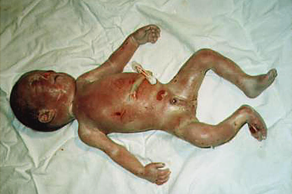
Almost everyone with this disorder has distinctive facial features, including a broad, flat nasal bridge and a high forehead. The eyes are widely spaced and may be protruding. Other characteristic facial features include a shortened distance between the nose and upper lip (a short philtrum), a downturned mouth, a small chin (micrognathia), and poorly formed ears with small holes (pits) or flaps of skin (tags). Additionally, affected individuals may have asymmetrical facial features and an unusually small head (microcephaly).
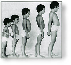
People with Wolf-Hirschhorn syndrome experience delayed growth and development. Slow growth begins before birth, and affected infants tend to have problems feeding and gaining weight (failure to thrive). They also have weak muscle tone (hypotonia) and underdeveloped muscles. Motor skills such as sitting, standing, and walking are significantly delayed. Most children and adults with this disorder also have short stature.
Intellectual disability ranges from mild to severe in people with Wolf-Hirschhorn syndrome. Compared to people with other forms of intellectual disability, their socialization skills are strong, while verbal communication and language skills tend to be weaker. Most affected children also have seizures, which may be resistant to treatment. Seizures tend to disappear with age.
Additional features of Wolf-Hirschhorn syndrome include skin changes such as mottled or dry skin, skeletal abnormalities such as abnormal curvature of the spine (scoliosis and kyphosis), dental problems including missing teeth, and an opening in the roof of the mouth (cleft palate) and/or in the lip (cleft lip). Wolf-Hirschhorn syndrome can also cause abnormalities of the eyes, heart, genitourinary tract, and brain.
A condition called Pitt-Rogers-Danks syndrome has features that overlap with those of Wolf-Hirschhorn syndrome. Researchers now recognize that these two conditions are actually part of a single syndrome with variable signs and symptoms.

It has been estimated that approximately 35% of individuals who have WHS die within the first two years of life. Many individuals who have WHS survive to adulthood. Universally, children with WHS have severe or profound developmental delays, however, there are many affected individuals who are able to walk and some that are able to talk in short sentences.

Frequency
The prevalence of Wolf-Hirschhorn syndrome is estimated to be 1 in 50,000 births. However, this may be an underestimate because it is likely that some affected individuals are never diagnosed.
For unknown reasons, Wolf-Hirschhorn syndrome occurs in about twice as many females as males.

Diagnosis
When WHS is suspected, chromosome analysis should be performed and the laboratory should be informed as to what syndrome is suspected. If the deletion is not visible, then fluorescent in situ hybridization (FISH) can be done specifically for the critical 4p16.3 region of chromosome 4.
Interestingly, there is a syndrome called Pitt-Rogers-Danks syndrome (PRDS) that has been reported to have similar characteristics to WHS. Several individuals who have initially been diagnosed with PRDS subsequently had FISH analysis that detected a deletion of 4p, and thus the individuals were reclassified as having WHS. Some feel that PRDS is actually WHS without obvious deletions of 4p.
When a couple has had a child diagnosed to have WHS, and a member of that couple carries a balanced translocation, genetic counseling should be offered to discuss reproductive options.
If ultrasound examination reveals findings consistent with the possibility of WHS in a family with no history of WHS, genetic counseling and prenatal diagnosis should be offered.


Causes
Wolf-Hirschorn is caused by a chromosomal deletion that occurs as a random (de novo) event during the formation of reproductive cells (eggs or sperm) or in early embryonic development. More complex chromosomal rearrangements can also occur as de novo events, which may help explain the variability in the condition's signs and symptoms. De novo chromosomal changes occur in people with no history of the disorder in their family.
A small percentage of all people with Wolf-Hirschhorn syndrome have the disorder as a result of an unusual chromosomal abnormality such as a ring chromosome 4. Ring chromosomes occur when a chromosome breaks in two places and the ends of the chromosome arms fuse together to form a circular structure. In the process, genes near the ends of the chromosome are lost.
In the remaining cases of Wolf-Hirschhorn syndrome, an affected individual inherits a copy of chromosome 4 with a deleted segment. In these cases, one of the individual's parents carries a chromosomal rearrangement between chromosome 4 and another chromosome. This rearrangement is called a balanced translocation. 
Between 85 and 90 percent of all cases of Wolf-Hirschhorn syndrome are not inherited.

Treatment
There is no treatment for the underlying condition of WHS. Treatment and management for patients who have WHS are specific to each individual. For example, some individuals who have WHS may have heart defects or a cleft lip and/or palate that may require surgery, while others may not. Therefore, there is no specific treatment for individuals who have WHS, rather, the treatment and management is geared toward that particular individual’s needs and is likely to include several medical specialists. Physiotherapy and occupational therapy are recommended. Some patients require physical aids, e.g. wheel chair, splints, hearing aids etc. Patients with congenital heart defects, clubfeet, and cryptorchidism have to be surgically treated. Those with seizures need  recurrent EEGs and antiepileptic drugs. Information about patients who have WHS has been compiled and provides a comprehensive look into the natural history of this condition and the needed management. The collection of this information has shown that many of these individuals may achieve more development than was previously believed possible.
The following management recommendations have been made by Drs. Battaglia and Carey @ http://wolfhirschhorn.org/about-wolf-hirschhorn-syndrome/:
  • Feeding problems should be addressed and may require interventionsuch as placement of a gastrostomy tube.
  • Characterization of seizures is important and treatment with antiepileptic medications such as valproic acid should be investigated and may help control the seizure activity in many individuals.
  • Skeletal abnormalities such as clubfoot should be addressed and treatment should be considered. It should not be assumed that clubfoot does not need addressed because the child will never walk. Children with WHS have learned to walk unassisted.
  • As approximately 30% of individuals may have congenital heart defects, the heart should be examined.
  • Hearing loss may occur and because some children are able to learn to talk in short sentences, they should be screened for hearing problems.
Eye abnormalities may be present and thus an ophthalmology exam should be performed to rule out any eye problems, even if no obvious signs are present.

Advices
 In regards to the development of patients with WHS, it is suggested that individuals participate in personal development programs to assist with social and occupational therapy for motor skills.

References
http://ghr.nlm.nih.gov/condition/wolf-hirschhorn-syndrome

Photos from:
https://blogger.googleusercontent.com/img/b/R29vZ2xl/AVvXsEjXrFTNKz9va7SlCZN7SwWQGlLC4dyQzml6SHy9fGhUNhOYuTvHVphDOP97vNPInVXAb-Lw3CUY8b_V5F3uaXkefi2B1hh-1ywWjuuYfRShIqHdoSsJasV2DBsabzze0ocyI-Q9Su8PTdyh/s1600/DSC_0033soft.jpg
http://upload.wikimedia.org/wikipedia/commons/f/ff/Wolf-hirschhorn.jpg

Phenylketonuria


Characteristics/Phenotype
Phenylketonuria is a rare metabolic condition in which a baby is born without the ability to properly break down an amino acid called phenylalanine. If not treated shortly after birth, PKU can be destructive to the nervous system, causing mental retardation.
Phenylketonuria (commonly known as PKU) is an inherited disorder that increases the levels of a substance called phenylalanine in the blood. Phenylalanine is a building block of proteins (an amino acid) that is obtained through the diet. It is found in all proteins and in some artificial sweeteners.
This condition is inherited in an autosomal recessive pattern, which means both copies of the gene in each cell have mutations. The parents of an individual with an autosomal recessive condition each carry one copy of the mutated gene, but they typically do not show signs and symptoms of the condition.
If PKU is not treated, phenylalanine can build up to harmful levels in the body, causing intellectual disability and other serious health problems. The signs and symptoms of PKU vary from mild to severe. The most severe form of this disorder is known as classic PKU. Infants with classic PKU appear normal until they are a few months old. 
Babies born with PKU usually have no symptoms at first. But if the disease is left untreated, babies experience severe brain damage. This damage can cause epilepsy, behavioral problems, and stunt the growth of the baby. Other symptoms include: eczema (skin rash), a musty body odor (from too much phenylalanine), a small head (microcephaly), and fair skin (because phenylalanine is necessary for skin pigmentation). Children with classic PKU tend to have lighter skin and hair than unaffected family members.
Less severe forms of this condition, sometimes called variant PKU and non-PKU hyperphenylalaninemia, have a smaller risk of brain damage. People with very mild cases may not require treatment with a low-phenylalanine diet.

Frequency
The occurrence of PKU varies among ethnic groups and geographic regions worldwide. In the United States, PKU occurs in 1 in 10,000 to 15,000 newborns. 

Diagnosis
Because PKU must be treated early, babies are routinely tested for the disease. Diagnosis is made through Newborn Screening. A small blood sample is taken from the baby's heel or arm and checked in a laboratory for high levels of phenylalanine.

Cause/s
PKU is caused by a mutation in a gene on chromosome 12. The gene codes for a protein called PAH (phenylalanine hydroxylase), an enzyme in the liver. This enzyme breaks down the amino acid phenylalanine into other products the body needs. When this gene is mutated, the shape of the PAH enzyme changes and it is unable to properly break down phenylalanine. Phenylalanine builds up in the blood and poisons nerve cells (neurons) in the brain.

Treatments/ Recommendations
People who have PKU must eat a protein-free diet, because nearly all proteins contain phenylalanine. Infants are given a special formula without phenylalanine. Older children and adults have to avoid protein-rich foods such as meat, eggs, cheese, and nuts. They must also avoid artificial sweeteners with aspertame, which contains phenylalanine.

References
http://health.nytimes.com/health/guides/disease/phenylketonuria/overview.html

Photo from: https://blogger.googleusercontent.com/img/b/R29vZ2xl/AVvXsEiB0CbaLQJvDdEY3-2fNCIddJsxl0DfHZiNiGYE2B2T5ZitnTwn7nKlzynxGpkqEHYsBiq7wx-tmKPNwS29xoCV0dSZIKMvKAHqfhQ-0Qf76JuIKt3L-veewnz2V9qxLmXu-lHEXB8ubWY/s400/images+(3).jpg

DUCHENNE MUSCULAR DYSTROPHY


Duchenne muscular dystrophy (DMD) is a rapidly progressive form of muscular dystrophy that occurs primarily in boys.

  • It is caused by an alteration (mutation) in a gene, called the DMD gene that can be    
           inherited in families in an X-linked recessive fashion, but it often occurs in people from 
           families without a known family history of the condition

  • Individuals who have DMD have progressive loss of muscle function and weakness, which 
            begins in the lower limbs. The DMD gene is the second largest gene to date, which encodes    
            the muscle protein, dystrophin.


Frequency:  1/3500 Male births


Phenotype/ Characteristics:

Duchenne muscular dystrophy is inherited in an X-linked recessive pattern. Males have only one copy of the X chromosome from their mother and one copy of the Y chromosome from their father. If their X chromosome has a DMD gene mutation, they will have Duchenne muscular dystrophy. Females, on the other hand, have two copies of the X chromosomes. Since females have two copies of this gene, if one copy does not work, they have a second back up copy to produce the dystrophin protein.


Understanding the inheritance of DMD!


Each son of a female carrier has a 50% chance of inheriting DMD through his mother’s faulty X chromosome and each daughter has a 50% chance of being a carrier of the disorder in the same way. Soon after the diagnosis of DMD it is essential that genetic counselling is arranged, together with appropriate tests for those members of the family who are at risk of being carriers. Genetic counselling provides information on the inheritance pattern, risks to other family members, and the ‘prognosis’ (likely outcome of the disorder). This service also provides information about diagnostic testing, including prenatal testing, as well as carrier testing.

What are the symptoms of Duchenne muscular dystrophy?

 First noticeable symptom is delay of motor milestones, including sitting and standing 
    independently.
> The mean age for walking in boys with Duchenne muscular dystrophy is 18 months.
> There is progressive muscle weakness of the legs and pelvic muscles, which is associated 
    with a loss of muscle mass (wasting).
 Calf muscles initially enlarge and the enlarged muscle tissue is eventually replaced with fat  
     and connective tissue (pseudohypertrophy).
 Symptoms usually appear in boys aged 1 to 6. There is a steady decline in muscle strength 
     between the ages of 6 and 11 years. By age 10, braces may be required for walking, and by
     age 12, most boys are confined to a wheelchair. Bones develop abnormally, causing 
     skeletal deformities of the spine and other areas.
 Muscular weakness and skeletal deformities frequently contribute to breathing disorders
> Cardiomyopathy (enlarged heart) occurs in almost all cases, beginning in the early teens in  
     some, and in all after the age of 18 years.
Intellectual impairment may occur, but it is not inevitable and does not worsen as the disorder 
    progresses.
> Few individuals with DMD live beyond their 30s. Breathing complications and     
    cardiomyopathy are common causes of death


Diagnosis:

Ø  A clinical diagnosis may be made when a boy has progressive symmetrical muscle weakness.

Ø  The symptoms present before age 5 years, and they often have extremely elevated creatine kinase blood levels (which are described below) .

Ø   If untreated, the affected boys become wheelchair dependent before age 13 years.

Ø  A muscle biopsy (taking a sample of muscle) for dystrophin studies can be done to look for abnormal levels of dystrophin in the muscle.

Ø  Genetic testing (looking at the body's genetic instructions) on a blood sample for changes in the DMD gene can help establish the diagnosis of Duchenne muscular dystrophy without performing a muscle biopsy.

Ø  For the remaining individuals, a combination of clinical findings, family history, blood creatine kinase concentration and muscle biopsy with dystrophin studies confirms the diagnosis.


Treatment:

Ø  Treatment for Duchenne muscular dystrophy is aimed at the symptoms.

Ø  Aggressive management of dilated cardiomyopathy with anti-congestive medications is used, including cardiac transplantation in severe cases.

Ø  Assistive devices for respiratory complications may be needed, especially at night.

Ø  The medication prednisone — a steroid — is given to improve the strength and function of individuals with DMD. Prednisone has been shown to prolong the ability to walk by 2 to 5 years. However, the possible side effects of prednisone include weight gain, high blood pressure, behavior changes, and delayed growth.

Ø   A synthetic form of prednisilone, called Deflazacort, is used in Europe and believed to have fewer side effects than prednisone.

Ø   A medication called cyclosporine has been used and has improved clinical function in children, but its use is controversial due to cyclosporine-induced myopathy.

Ø  Oxandrolone, a medication used in a research study, has similar effects to prednisone with fewer side effects. 


Recommendations/advices:

--> Communication is a key word in the coping process. If parents can speak openly and honestly with each other, sharing concerns and discussing ways to meet challenges, the whole family will benefit. 

-->  Be open and approachable. Encourage him to discuss his needs, to learn to ask others for assistance when he needs it and to politely refuse when he can manage independently.

-->  A healthy diet, with restriction on foods containing high levels of fat and sugar, and including plenty of fluids, fruit and vegetables can be encouraged.

--> Encourage special interests, physical exercise such as swimming, according to his mobility, and relationships with peers.

-->  Always remember that he needs support, love, a warm and caring family environment and not least, the encouragement to live life as fully as possible with his friends and family.

--> Rather than leaving the majority of the additional responsibilities to one family member, it is advisable to involve all family members as much as possible.

 --> Creating new friendships with other DMD families (or families living with other disabilities) can be rewarding for all concerned


References:

Learning About Duchenne Muscular Dystrophy (2010). Retrieved on March 12, 2013 from http://www.genome.gov/19518854

Duchenne Muscular Dystrophy (2011). Retrieved on March 12, 2013 from http://www.umm.edu/ency/article/000705.htm

Photos from:
http://www.loyarburok.com/wp-content/uploads/2011/05/Duchenne.jpg
https://blogger.googleusercontent.com/img/b/R29vZ2xl/AVvXsEipgwK6ctNeTnOeaP6BQbMWKLvEXpg9LH56jH5P8ptfuP08GzM9zBqvXPH168QTO_ubFaaJDl8iXeXet6M42HSAQDK_Sir4tJyQGrylrN0AdfR7aVxqRsnU9VgFZNKIKObE5yDNyStZwnc/s400/duchenne.gif
https://blogger.googleusercontent.com/img/b/R29vZ2xl/AVvXsEiolPlWzfaukruqRDGvTlTtLpEFMTLt3XIgDVPBfn_xq3zhQoYZfNzII63Y0uwlBKmUtjrcrp5fS4GcCbLf_IDT5rJ7BumcYIMVP1FDaQh6bOn9SdbNWw186fJaZax08AsEJkRq0mcrOag/s320/d.jpg


Jacob's Syndrome


Characteristics/Phenotype
Jacobs syndrome is a rare chromosomal disorder that affects only males. It is a condition where there is the presence of an extra Y chromosome in all of a male's cells. Males normally have only one X and one Y chromosome, however, individuals with Jacob's syndrome have at least one X and at least two Y chromosomes (XYY). It gives males a total of 47 chromosomes, instead of the average of 46.
Other names for Jacobs syndrome:
  • 47, XYY syndrome
  • XYY Karyotype
  • XYY syndrome
  • YY syndrome
  • Diplo-Y Syndrome
  • Polysomy Y
  • Super-male syndrome
This condition is usually asymptomatic which means that it shows no evidence of disease.
In children, 47,XYY may produce some of the following symptoms:
• Delay in developing speech
• Low muscle tone (hypotonia)
• Delayed social development
• Learning disabilities
• Pervasive developmental disorder (PDD-NOS) or mild autism spectrum disorder
• Attention deficit and/or hyperactivity
In older teens and adults, 47,XXY may also be characterized by:
• Very tall stature and long limbs
• Severe acne
• Continued learning and/or social difficulties
• Delay in vocational success
• Anxiety, depression or other mood disorder
• Impulsivity
• Dental problems

Frequency
This condition occurs in about 1 in 1,000 newborn boys. Five to 10 boys with 47,XYY syndrome are born in the United States each day.

Diagnosis
Diagnosis requires a specialized blood test, called a karyotype or a buccal swab test called
XCAT. In some cases, FISH (fluorescence in situ hybridization), or microarray analysis is performed.
Prenatal diagnosis requires amniocentesis or chorionic villus sampling, or a noninvasive prenatal test called NIPT.

Cause/s
Most cases of 47,XYY syndrome are not inherited. The chromosomal change usually occurs as a random event during the formation of sperm cells. An error in cell division called nondisjunction can result in sperm cells with an extra copy of the Y chromosome. If one of these atypical reproductive cells contributes to the genetic makeup of a child, the child will have an extra Y chromosome in each of the body's cells.
46,XY/47,XYY mosaicism is also not inherited. It occurs as a random event during cell division in early embryonic development. As a result, some of an affected person's cells have one X chromosome and one Y chromosome (46,XY), and other cells have one X chromosome and two Y chromosomes (47,XYY).

Treatments
Most symptoms can be addressed successfully by early diagnosis and detection, appropriate treatments such as speech and occupational therapy, and special education services, if learning disabilities are involved. Infants and very young children may be eligible for early intervention. Children can benefit from special
education services including;
• Speech therapy
• Occupational and physical therapy when indicated for motor problems
• Special education services or educational accommodations when necessary
• Social skills training programs
• Family or individual counseling
• Behavioral consultation
• Regular exercise and upper body strengthening programs
Adolescents with 47,XYY, may develop severe acne. If so, they should be seen by a dermatologist and treated appropriately to prevent scarring. Adults with 47,XYY, may take several years longer to develop the emotional maturity to become self-supporting and capable of living independently. Interventions that may be helpful for adults with XYY include:
• Psychological counseling when indicated
• Vocational counseling and workplace accommodations when necessary
• Dental health preservation/restoration
• A healthy diet and regular exercise to maintain cardiovascular health

Recommendations/Advices
As a parent, tell your child. . .

References
http://www.mamashealth.com/syndrome/jacob.asp

Photos from:
https://blogger.googleusercontent.com/img/b/R29vZ2xl/AVvXsEjehaVzLJ74kuJH07jDuTDMsNhBvvb3SnHOdKvKgQoKXgQ71sV5to6Ge0EjCf1ELcU12m5eU70ixxRXspUZAsg-8mUK3_cr4LKPP6kYMy3y393FB960_wf4WZP6fiRVhZlQzzFwhBwNdiA/s400/images+(2).jpg
https://blogger.googleusercontent.com/img/b/R29vZ2xl/AVvXsEgu4mE8uo91XW7warqrPRNQtxwMH8MAYxTMyN3biOm3f7D3FW7XVU5eFG8JoTCI-FgPtdWm2kCzqNz4rcUDcyVsiHRUH_ycywbd9IMgMRcVRSkMlMoPZH1kbGqeByk_ecfkhExdECG3-3k/s320/xyykaryotype.jpg
http://www.blogger.com/blogger.g?blogID=954453551672198011#editor/target=post;postID=5843684501468290769

PATAU SYNDROME


> Patau syndrome is the least common and most severe of the viable autosomal trisomies.
> Median survival is fewer than 3 day
> First identified as a cytogenetic syndrome in 1960, Patau syndrome is caused by an extra  
   copy of     chromosome 13, a medium-length acrocentric chromosome. 
> The extra copy of chromosome 13 in Patau syndrome causes severe neurological and heart 
     defects which       make it difficult for infants to survive. 
> Patau syndrome appears to affect females more than males, most likely because male    
    fetuses do not        survive until birth.
> Patau syndrome, like Down syndrome, is associated with increased age of the mother. It may 
   affect  individuals of all ethnic backgrounds.

Frequency of Births: 1/15000


Characteristics:


Causes:

>  The cause of the trisomy in Patau and Down syndromes is similar. A phenomenon called nondisjunction is responsible for most cases of trisomy 13 and 21. 
> Rather than splitting evenly, cells that have undergone nondisjunction typically split unevently into two cells; one cell receives one extra copy of a chromosome, while the other is deficient in the chromosome.

> A major risk factor for nondisjunction is advanced maternal age. The chance of nondisjunction increases significantly once mothers are 35 years old and greater. As you'd expect, the risk of Patau and Down syndromes increases considerably with advanced maternal age.

Diagnosis: 

The symptoms of Patau syndrome are evident at birth. Infants with Patau syndrome may present with a single umbilical artery at birth. Many times there are signs of congenital heart disease, such as atrial septal defect, abnormal placement of their heart towards the ride side of their chest, ventricular septal defect, or patent ductus arteriosus. Ultrasound or gastrointestinal X-rays might display rotation of the infant's internal organs.
Imaging studies such as computed tomography (CT) or magnetic resonance imaging (MRI) should be done to look for brain, heart, and kidney defects. An ultrasound of the heart (echocardiogram) should be done given the high frequency of heart defects associated with Patau syndrome. 

Treatment


Some infants born with Patau syndrome have severe and incurable birth defects. However, children with better prognoses require medical treatment to correct structural abnormalities and associated complications. Treatment of children with Patau syndrome involves planning on a case-by-case basis. The forms of treatment a particular person receives is dependent upon the person's particular condition. Intervention through surgical means are commonly withheld for the first few months of the person's life due to the high mortality rate of children with Patau syndrome.

Prognosis:
Approximately 45% of trisomy 13 babies die within their first month of life; up to 70% in the first six months; and over 70% by one year of age. Survival to adulthood is very rare. Only one adult is known to have survived to age 33.
Most survivors have profound mental and physical disabilities; however, the capacity for learning in children with Patau syndrome varies from case to case. Older children may be able to walk with or without a walker. They may also be able to understand words and phrases, follow simple commands, use a few words or signs, and recognize and interact with others.

Most survivors have profound mental and physical disabilities; however, the capacity for learning in children with Patau syndrome varies from case to case. Older children may be able to walk with or without a walker. They may also be able to understand words and phrases, follow simple commands, use a few words or signs, and recognize and interact with others. 

References:

Best, R., Buehler, B. (2012). Patau Syndrome. Retrieved on March 12, 2013 from http://emedicine.medscape.com/article/947706-overview.

Kugler, M. (2005). Patau Syndrome (Trisomy 13). Retrieved on March 12, 2013 from http://rarediseases.about.com/od/rarediseasesp/a/patau05.htm.

Photos from:
https://blogger.googleusercontent.com/img/b/R29vZ2xl/AVvXsEhtMPLrJkmR7i6W39vDSGRbOlBG4AoZdkRi62itDQV3GNIpgjuo2-lC1fSv9cwMq2s7O_5SXjSGHrUdYLhcrjvTSak5FY38IhHDq0ttWC4hQakNSMsgq-5Fu-UE68ydBN3dl2WzoMMQXU4/s400/pat.gif
https://blogger.googleusercontent.com/img/b/R29vZ2xl/AVvXsEg2WnNkADdVICiUuwBn6q7G4mF2HaFTWdohN9NHczf-fLkuGIzbRqe5ceaN1a8Ou5uSwwTlpKP6CoO_389uygPO0MwO7ZI_h-TpzbC9cBlTh2IQdlAvxfAl5ce2nNZKMHa7oNBcPZr8-Sw/s320/patau.jpg