Mga Pahina

Martes, Marso 12, 2013

ANGELMAN SYNDROME


Overview: Angelman syndrome is a neuro-genetic disorder, caused by genomic imprinting. The deletion or inactivation of genes on the maternally inherited chromosome 15 while the paternal copy, which may be of normal sequence, is imprinted and therefore silenced causes the condition. It is the sister syndrome of Prader-Willi syndrome.


Phenotype/Characteristics: A patient with Angelman syndrome will have the following signs and symptoms:
  •      a severe delay in development from 6-12 months
  •      speech problems (most children do not speak or only say a few words)
  •      movement or balance problems – usually ataxia (loss of co-ordination) and jerky movements of the limbs

A child with Angelman syndrome will also display distinctive behaviours, these may include:
  •      frequent laughter and smiling, with little stimulus
  •      being easily excitable, often flapping the hands
  •      being restless
  •      having a short attention span

Many children with Angelman syndrome will have microcephaly by the age of two, and many will develop seizures around this age.

Some children with Angelman syndrome will also have the following features and behaviours:
  •      a flattened skull at the back
  •      a tendency to stick out the tongue
  •      feeding problems during infancy
  •      a prominent chin
  •      a wide mouth and wide-spaced teeth
  •      frequent drooling
  •      excessive chewing
  •      strabismus
  •      pale skin and light hair and eye colour
  •      uplifted bent arms, especially during walking
  •      increased sensitivity to heat
  •      disturbed sleep, and needing less sleep than usual
  •      attraction to, or fascination with, water
  •      scoliosis


Frequency: Angelman syndrome affects an estimated 1 in 12,000 to 20,000 people.
Diagnosis: A deletion or inactivity on chromosome 15 by array comparative genomic hybridization (aCGH) or by BACs-on-Beads technology can be a diagnostic method. FISH-metaphase or interphase, sequence analysis of the coding region and the like may also be used. Prenatal diagnosis is also feasible.

Causes:  The condition is caused by the malfunction of the UBE3A gene on chromosome number 15. Normally, both parental copies of the gene are active in the body’s tissues. In certain areas of the brain, only the gene inherited from the mother is active. If the maternal copy of the UBE3A gene is lost because of a chromosomal change or a gene mutation, a person will have no active copies of the gene in some parts of the brain.
In most cases of Angelman syndrome, the child's maternal copy of the UBE3A gene is not functioning normally or is missing altogether.
Several different genetic mechanisms can inactivate or delete the maternal copy of the UBE3A gene. Most cases of Angelman syndrome (about 70 percent) occur when a segment of the maternal chromosome 15 containing this gene is deleted. In other cases (about 11 percent), Angelman syndrome is caused by a mutation in the maternal copy of the UBE3A gene.
In a small percentage of cases, Angelman syndrome results when a person inherits two copies of chromosome 15 from his or her father (paternal copies) instead of one copy from each parent (paternal uniparental disomy). Rarely, Angelman syndrome can also be caused by a chromosomal rearrangement called a translocation, or by a mutation or other defect in the region of DNA that controls activation of the UBE3A gene. These genetic changes can abnormally turn off (inactivate) UBE3A or other genes on the maternal copy of chromosome 15.


Treatment/Recommendations/Therapies: There is no cure for Angelman syndrome and some of the symptoms can be difficult to manage.
The following treatments and aids may augment the effects of the condition to a certain extent:
  •      Anti-epileptic medicine to control the seizures –sodium valproate, clonazepam and ethosuccimide are some of the drugs in common use.
  •      Physiotherapy, attention to their sitting posture, a back brace and possibly spinal surgery to prevent their spine from curving further (preventing scoliosis). 
  •      A feeding tube and other feeding aids, especially in infancy. In older children, weight gain may be a problem.
  •      Occupational therapy to help them develop skills needed for dressing, feeding and other daily living activities.
  •      An ankle foot orthosis (lower leg brace) to help them walk independently.
  •      Picture boards, eye gaze technology and other visual aids to help them communicate.

Therapeutic horseriding, swimming, hydrotherapy and music therapy have been reported to be beneficial.

Additional Fact: In the 2011 Philippine drama series Budoy, the titular character and main protagonist Budoy Maniego (played by Filipino actor Gerald Anderson) is diagnosed with Angelman Syndrome.

Support Groups:
Angelman syndrome foundation
http://www.angelman.org/


References:

NHS. (2012). Angelman syndrome. NHS. Retrieved March 12, 2013 from the URL: http://www.nhs.uk/conditions/angelman-syndrome/Pages/Introduction.aspx

Genetics Home reference (2012). Angelman Syndrome. Retrieved March 12, 2013 from the URL: http://ghr.nlm.nih.gov/condition/angelman-syndrome

 Photo from:

http://www.angelman.org/_angelman/cache/file/29CCDE00-6C72-4EC0-BFCCF69C7E2EC9B7.jpg

PRADER WILLI SYNDROME


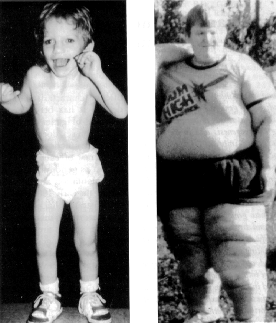
Overview: Prader-Willi syndrome (PWS) is a disorder caused by a deletion or disruption of genes in the proximal arm of chromosome 15 or by maternal disomy in the proximal arm of chromosome 15.

Patient with Prader Willi Syndrome


Phenotype/Characteristics and Diagnosis:
A diagnostic criteria for Prader-Willi syndrome was made by Holm et al. Based on these guidelines, the diagnosis of Prader-Willi syndrome is highly likely in children younger than 3 years with 5 points (3 from major criteria) or in those older than 3 years with 8 points (4 from major criteria).
·         Major criteria (1 point each)
o    CNS - Infantile central hypotonia
o    GI - Infantile feeding problems and/or failure to thrive
o    Nutrition - Rapid weight gain in children aged 1-6 years
o    Craniofacial - Characteristic facial features such as narrow bifrontal diameter, almond-shaped palpebral fissures, narrow nasal bridge, and down-turned mouth
o    Endocrine - Hypogonadism
o    Developmental - Developmental delay and/or mental retardation
·         Minor criteria (one half point each)
o    Neurologic - Decreased fetal movement and/or infantile lethargy
o    Pulmonary - Sleep disturbance and/or sleep apnea
o    Endocrine - Short stature for predicted height by mid adolescence
o    Dermatologic - Hypopigmentation
o    Orthopedic - Small hands and feet
o    Orthopedic - Narrow hands with straight ulnar border
o    Ophthalmologic – Esotropia and/or myopia
o    Dental - Thick viscous saliva
o    Otolaryngology - Speech articulation defects
o    Psychiatric - Skin picking (Some patients with Prader-Willi syndrome have become anemic from chronic rectal bleeding secondary to skin picking.)
·         Supportive criteria (no points)
o    Neurology - High pain threshold and normal neuromuscular evaluation for hypotonia
o    Gastroenterology - Decreased vomiting
o    Endocrinology - Ineffective thermoregulation, early adrenarche, and/or osteoporosis, adrenal insufficiency
o    Orthopedics – Scoliosis or kyphosis
o    Developmental - Jigsaw puzzle proficiency

Frequency: Prader-Willi syndrome affects an estimated 1 in 10,000 to 30,000 people worldwide. Differences in prevalence rates between sexes have not been reported

Diagnosis:
Genetic testing is employed for accurate diagnosis of the condition.
· Genetic testing for Prader-Willi syndrome (PWS) includes chromosomal analysis and assessment for methylation patterns in the Prader-Willi syndrome region.
· Methylation patterns can be determined with Southern blot hybridization or polymerase chain reaction (PCR) using DNA primers that can detect methylated cytosine.
· Analysis for underlying uniparental disomy requires samples from both parents and the child with Prader-Willi syndrome.
· Fluorescent in situ hybridization (FISH) can be used to confirm prenatal diagnosis when a deletion in the 15q region is suspected after chorionic villus sampling or amniocentesis.

Causes:
·     Prader-Willi syndrome is due to the loss of the paternal copy of chromosome 15q11.2-13.
·     Most cases of Prader-Willi syndrome are sporadic. More than 70% of patients have a deletion of the paternal copy; approximately 25% of patients with Prader-Willi syndrome have maternal uniparental disomy in chromosome 15. The remainder of patients with this disorder have a translocation or other structural alteration in chromosome 15.
·     Most manifestations of Prader-Willi syndrome are attributable to hypothalamic dysfunction.

Treatment/Recommendations/Therapies:
Patients with Prader-Willi syndrome (PWS) frequently require medical care for the following:[37]
·         Initial management of hypotonia or poor feeding
·         Evaluation for hypogonadism or hypopituitarism
·         Management of obesity
·         Monitoring for scoliosis
·         Therapy for behavioral issues

Patients with Prader-Willi syndrome may require surgical care for treatment of complications of obesity, treatment of cryptorchidism, and scoliosis intervention. 

Tonsillectomy, adenoidectomy, or tracheostomy placement may be required in patients with obstructive sleep apnea.

Biliopancreatic diversion and gastric bypass surgery have been ineffective for long-term weight reduction. Patients with Prader-Willi syndrome have hyperphagia and require restricted access to foods to minimize weight gain. Binge-eating episodes may predispose patients to development of food poisoning and acute gastric dilation. Caregivers of patients with Prader-Willi syndrome should be instructed in the Heimlich maneuver.

Growth hormone therapy in patients with growth hormone deficiency improves lean body mass and corrects osteopenia.

Patients with Prader-Willi syndrome (PWS) may require inpatient evaluation and treatment for hypotonia and poor feeding during infancy.

Individuals with Prader-Willi syndrome and other medical issues, including scoliosis and complications of obesity or pickwickian syndrome, may require inpatient therapy.

Patients with severe behavioral problems may merit admission to a facility staffed with individuals with long-term experience with Prader-Willi syndrome.

Support Groups:

Support - Prader-Willi Syndrome Association

http://www.pwsausa.org/support/


References:

A.D.A.M.Health Solutions, Ebix, Inc. (2012) Prader-Willi syndrome. NCBI. Retrieved March 12, 2013 from the URL: http://www.ncbi.nlm.nih.gov/pubmedhealth/PMH0002572/

Genetics Home Reference (2011). Prader Willi Syndrome. Genetics Home reference. Retrieved March 12, 2013 from the URL: http://ghr.nlm.nih.gov/condition/prader-willi-syndrome
Scheimann, A. (2012). Prader-Willi syndrome. Medscape Reference. Retrieved March 12, 2013 from the URL: http://emedicine.medscape.com/article/947954-overview

Photo from:
http://schoolworkhelper.net/wp-content/uploads/2011/06/Prader-Willi-Syndrome-2.gif


Turner's Syndrome


Characteristics/Phenotypes


Turner's syndrome is a genetic disorder that affects females. Usually, a female has two X chromosomes; in females with Turner's syndrome, one of these chromosomes is missing or abnormal. Other names for Turner's syndrome include monosomy X, 45X and Ullrich-Turner syndrome.
The characteristic of the disease is mainly depending on the age of the girl or woman affected.
 Babies born with Turner syndrome often have swollen hands and feet caused by lymphoedema. Prominent thick neck tissue, swelling of the neck (cystic hygroma) and being a small baby.
The two most common characteristics of Turner syndrome are:
  • short height
  • ovaries that do not function
These two characteristics affect more than nine out of ten females with Turner syndrome.  Babies with Turner syndrome are usually a normal height until the age of three. After this age, they will have a lower-than-average growth rate.
At puberty, a girl with Turner syndrome will not have the normal growth spurt. Puberty usually happens in girls aged between eight and thirteen. Without treatment, someone with Turner syndrome will usually grow to around 136-147cm (4ft 6in to 4ft 10in). On average, adult women with untreated Turner syndrome are 20cm (8in) shorter than adult women without Turner syndrome. Treatment with growth hormones can reduce this difference.
Ovaries are the pair of reproductive organs in females that produce eggs and sex hormones. During puberty, a girl's ovaries usually begin to produce the sex hormones oestrogen and progesterone. However, most girls who have Turner syndrome will not produce these sex hormones, which means that they:
  • may not start their periods naturally as other girls do  
  • may not fully develop breasts
  • may be infertile (unable to conceive a baby)
As well as short height and non-functioning ovaries, there are many other symptoms or characteristics that can affect girls and women with Turner syndrome. Some of these are listed below.
Symptoms that affect appearance include: 
  • a particularly short, wide neck (webbed neck)
  • a broad chest and widely spaced nipples
  • arms that turn out slightly at the elbows
  • a low hairline
  • abnormalities of the mouth, which can cause problems with the teeth
  • a large number of moles
  • small, spoon-shaped nails 
  • a short fourth finger or toe
  • eyes that slant downwards
  • an eye problem (cataract, ptosis)
  • low-set ears 
  • hearing loss, which can develop in later life, is often more severe and develops earlier than the normal age-related decline in hearing
Most girls with Turner syndrome have good language and reading skills. However, some have behavioural, social and specific learning difficulties that are often overlooked by doctors that can cause great anxiety to the parents. They include: 
  • problems with social intelligence 
  • problems with spatial awareness and numeracy
  • attention and hyperactivity problems

Frequency
This condition occurs in about 1 in 2,000-2,500 female births worldwide, but is much more common among pregnancies that do not survive to term (miscarriages and stillbirths).

Diagnosis

The condition may be diagnosed at various life stages including:
·         Before birth (prenatally) – usually if an amniocentesis has been performed or abnormalities are seen during an ultrasound
·         At birth – due to certain physical features
·         In childhood – when the young girl doesn’t grow at a similar rate to her peers
·         During the teenage years – when puberty fails to arrive
·         In adulthood – during investigations for infertility.
Turner’s syndrome is diagnosed using a number of tests including:
·         Amniocentesis and chorionic villus sampling (before birth)
·         Karyotyping
·         Clinical history
·         Physical examination
·         Psychological and educational assessment
·         Blood tests and chromosome analysis Genetic tests.

Cause/s

Turner syndrome is typically caused by what is called nondisjunction. If a pair of sex chromosomes fails to separate during the formation of an egg (or sperm), this is referred to as nondisjunction. When an abnormal egg unites with a normal sperm to form an embryo, that embryo may end up missing one of the sex chromosomes (X rather than XX). As the embryo grows and the cells divide, every cell of the baby's body will be missing one of the X chromosomes.

The abnormality is not inherited from an affected parent (not passed down from parent to child) because women with Turner syndrome are usually sterile and cannot have children.
In about 20 percent of Turner syndrome cases, one X chromosome is abnormal. It may be shaped like a ring, or missing some genetic material.

About 30 percent of girls with the disorder are only missing the X chromosome in some of their cells. This mixed chromosome pattern is known as mosaicism. Girls with this pattern may have fewer symptoms because they still have some normal (XX) cells.

Treatments/Therapies


Hormone replacement therapy is the best way to treat this disorder. Teenagers are treated with growth hormone to help them reach a normal height. They may also be given low doses of androgens (male hormones which females also produce in small quantities) to increase height and encourage normal hair and muscle growth. Some patients may take the female hormone estrogen to promote normal sexual development.
Babies born with a heart murmur or narrowing of the aorta may need surgery to correct the problem. A heart expert (cardiologist) will assess and follow up any treatment necessary.
Girls who have Turner syndrome are more likely to get middle ear infections. Repeated infections may lead to hearing loss and should be evaluated by the pediatrician. An ear, nose and throat specialist (ENT) may be involved in caring for this health issue. Almost all women are infertile, but pregnancy with donor embryos may be possible.

Recommendations/Advices


Remember that Regular health checks are very important.
Special clinics for the care of girls and women who have Turner syndrome are available in some areas, with access to a variety of specialists.
Early preventive care and treatment is very important.
Having appropriate medical treatment and support allows a woman with Turner syndrome to lead a normal, healthy and happy life.

References


http://www.betterhealth.vic.gov.au/bhcv2/bhcarticles.nsf/pages/Turner's_syndrome

Photos from:
https://blogger.googleusercontent.com/img/b/R29vZ2xl/AVvXsEgbOGxLpB7yrAeTjWHu5TioPgKTcJ8VCjSrK_0IiLl2S5OkoJyggolg3x5F0tzuhtVOOCQ4r4mtTQPY7zLPwsyFCRBluopFpoccE1gHfx8r68p1f0OvCKBExrdzpye-Ugok_0UWiM9H3SQ/s320/images+(1).jpg
https://blogger.googleusercontent.com/img/b/R29vZ2xl/AVvXsEiV2tLe1GJ2eUP3NNcYb74FpbGb9JzeTB8WKQ5lUvay2NvvVEjHDp-LuU3yynXSAwWFYdQmioFpkqJJY3nVC5OoT4pkw8kjNrM0lG5NJUr9XlmUvcdd25W7GNUfdVBZIL7S4zTh79v5fJ0/s320/X2604-T-53.png

CRYPTORCHIDISM


Alternate Names: Undescended testicles


Overview: Cryptorchidism literally means hidden or obscure testis and generally refers to an undescended or maldescended testis. Untreated cryptorchidism clearly has deleterious effects on the testis over time. 

Phenotype/Characteristics: Testicles absent from the normal scrotal position can be:
1. from high the posterior (retroperitoneal) abdomen, just below the kidney, to the inguinal ring;
2. in the inguinal canal;
3. ectopic, usually outside the inguinal canal and sometimes even under the skin of the thigh, the perineum, the opposite scrotum, or the femoral canal;
4. found to be undeveloped (hypoplastic) or severely abnormal (dysgenetic);
5. found to have vanished (anorchia)

The condition is associated with reduced fertility, increased risk of testicular tumors, and psychological problems. It also predisposes the patient to testicular torsion and inguinal hernias.

Frequency: About 3% of full-term and 30% of premature infant boys are born with at least one undescended testis. However, about 80% of cryptorchid testes descend by the first year of life (the majority within three months), making the true incidence of cryptorchidism around 1% overall.

Cryptorchidism is identified in 1.5-4% of fathers and 6.2% of brothers of patients with cryptorchidism. Heritability in first-degree male relatives is estimated to be 0.67.

Diagnosis: An examination confirms that one or both of the testicles are not in the scrotum.
The health care provider may or may not be able to feel the undescended testicle in the abdominal wall above the scrotum. Approximately 20-30% of patients with cryptorchidism have nonpalpable testes. Nonpalpable testes may be intra-abdominal or absent. Palpable testes may be undescended, ectopic, or retractile.

Causes: Cryptorchidism is multifactorial. Extensive research and clinical observations have elucidated some of the factors involved, but the exact mechanism of cryptorchidism has proven to be elusive.
Low birth weight often results in the condition.
Transabdominal descent of the testis involves differential growth of vertebrae and pelvis and is facilitated by the development of the gubernaculum, processus vaginalis, spermatic vessels, and scrotum. A normal hypothalamic-pituitary-gonadal axis is a prerequisite for testicular descent. Furthermore, testosterone and its conversion to dihydrotestosterone (DHT) are also necessary for continued migration, especially during the inguinoscrotal phase.
Although its exact mechanism of action is unclear, the gubernaculum has significant importance in undescended testes. In patients with cryptorchidism, the gubernaculum is not firmly attached to the scrotum, and the testis is not pulled into the scrotum. 
Intra-abdominal pressure also appears to play a role in testicular descent. Conditions associated with decreased pressure include prune belly syndrome,cloacal exstrophy, omphalocele, and gastroschisis, among other various syndromes. Each is associated with an increased risk of undescended testes.
Associated anomalies: Associated anomalies and conditions may include the following:
  •      Patent processus vaginalis
  •      Abnormal epididymis
  •      Cerebral palsy
  •      Mental retardation
  •      Wilms tumor
  •      Abdominal wall defects (eg, gastroschisis, omphalocele, prune belly syndrome)
  •      Hypospadias
Treatment/Recommendations/Therapies: Most cases get better on their own, without any treatment. Medicine or surgery to correct the condition is usually successful.
Cryptorchidism should be treated when the patient is aged approximately 6 months. 
Human chorionic gonadtropin (hCG) is administered via intramuscular injection and stimulate androgen production and testicular descent (mechanism still unknown). Ginadtropin releasing hormone promoted the same effect and may be used for a combined hormonal treatment.

Successful surgical placement of the testis in the scrotum (adequate mobilization of the testis and spermatic vessels, ligation of the associated hernia sac, and adequate fixation of the testis in a dependent portion of the scrotum) can be employed.

Support Groups:

I Have Cryptorchidism


References:

Kaneshiro, N. (2012) Undescended testicle. ADAM Medical Encyclopedia. Retrieved March 12, 2013 from the URL: http://www.ncbi.nlm.nih.gov/pubmedhealth/PMH0001968/

Sumfest, J. (2012). Cryptorchidism. Medscape reference. Retrieved March 12, 2013 from the URL: http://emedicine.medscape.com/article/438378-overview

Photo from:
http://wikis.lib.ncsu.edu/images/f/f6/Cryptorchidism.jpg